Oligodendroglia
 Myelin sheath |
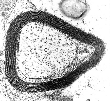
 Oligodendroglial cell |
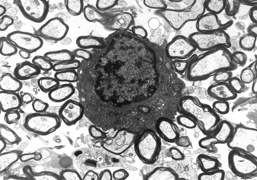
 Oligodendroglial cells |
Myelin is a sheath of plasma membrane wrapped around an axon. Its insulating properties are important for conductivity. It consists of 70-80% lipids and 20% structural proteins, including Proteolipid Protein, Myelin Basic Protein(MBP) and Myelin Associated Glycoprotein. In the CNS, myelin is made by oligodendroglial cells; in peripheral nerves by Schwann cells. Each Schwann cell makes one segment of myelin; each oligodendrocyte makes multiple segments of myelin that wrap around several axons.
Oligodendroglial cells are present in gray matter and in white matter. In paraffin sections, oligodendroglial cells have clear cytoplasm, an appearance referred to as "fried egg". This is an artefact of histological processing and is replicated in tumors of oligodendroglial cells.
Oligodendroglial cells are present in gray matter and in white matter. In paraffin sections, oligodendroglial cells have clear cytoplasm, an appearance referred to as "fried egg". This is an artefact of histological processing and is replicated in tumors of oligodendroglial cells.
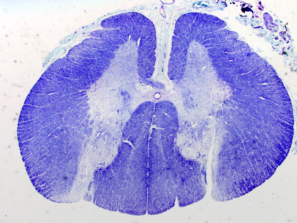
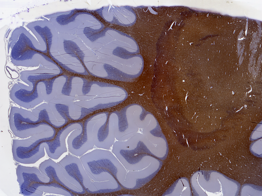
Myelin stain> |
Myelin basic protein immunostain> |
Most myelin stains use hematoxylin-based solutions that attach to phospholipid components of the myelin sheath and give myelin a deep blue color. Alternatively, immunostains with antibodies to MBP can be used to evaluate myelin in issue sections.
Myelin and oligodendrocytes are non-specifically lost in cerebral infarcts, infections, and other lesions that involve the white matter. They are especially sensitive to hypoxia and ischemia in premature infants. Oligodendrocyte injury in the perinatal period can cause white matter infarcts or permanent white matter loss around the lateral ventricles (periventricular leukomalacia). Oligodendrocyte loss occurs also in demyelinative disorders, such as multiple sclerosis and progressive multifocal leukoencephalopathy (PML). PML is caused by a papovavirus infection of oligodendrocytes. Myelin proteins are immunogenic, and some aspects of inflammatory demyelinative disorders are due to autoimmunity. Leukodystrophies are metabolic disorders caused by biochemical abnormalities of myelin lipids and proteins. They show diffuse and progressive loss of myelin. The term "degeneration" in reference to myelin means loss of myelin; in reference to the white matter or specific tracts (e.g. posterior colum degeneration), it means loss of myelin and axons.
In addition to myelinating oligodendrocytes, the mature brain contains a large number of oligodendrocyte precursor cells (OPCs), that can be distinguised by specific markers they express. OPCs are generated in fetal life and postnatally; they do not make myelin but retain their undifferentiated stem cell status. The potential of OPCs to differentiate and repair multiple sclerosis and other demyelinating disease lesions remains to be explored.
Ependyma
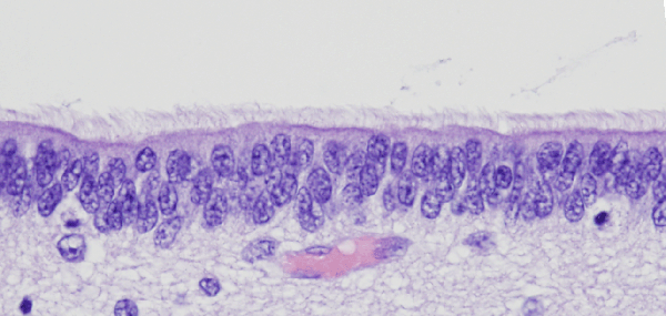 Ependyma |
The ependymal cells line the walls of the ventricles and form the specialized choroid plexus epithelium which secretes the cerebrospinal fluid (CSF). The ependymal lining may be injured and lost as a result of infections involving the cerebral ventricles (ventriculitis) and intraventricular hemorrhage. Residual ependymal cells proliferate and form tubules (rosettes) and small nodules with admixed reactive astrocytes (ependymal granulations) in the walls of the ventricles.
Further Reading
- Chong SY, Chan JR. Tapping into the glial reservoir: cells committed to remaining uncommitted. J Cell Biol 2010; 188: 305-12. PubMed