Question 2 : A 36 week gestation fetus is more susceptible to HIE than a 40 year old person
Correct A difficult question. Brain damage in HIE is mediated by glutamate, therefore glutamate receptors.
A 40 year old individual has more synapses than a 36 week fetus, hence is more vulnerable to glutamate mediated damage.
On the other hand, the brain of a 36 week fetus is larger in proportion to the body, and needs additional energy for growth.
Incorrect. A difficult question. Brain damage in HIE is mediated by glutamate, therefore glutamate receptors.
A 40 year old individual has more synapses than a 36 week fetus, hence is more vulnerable to glutamate mediated damage.
On the other hand, the brain of a 36 week fetus is larger in proportion to the body, and needs additional energy for growth.
Question 3: Porencephaly is a developmental malformation occurring in the second trimester
Incorrect Porencephaly is a destructive, probably ischemic lesion which occurs
in the second or third trimester.
Correct. Porencephaly is a destructive, probably ischemic lesion which occurs
in the second or third trimester.
Question 4: The pathology illustrated below is a developmental malformation occurring in the second trimester
.
False The lesion is porencephaly (a cavity in the brain and enlarged ventricle on the left),
which is not a developmental malformation but a destructive, probably ischemic lesion which occurs in the second or third trimester.
True The lesion is porencephaly (a cavity in the brain and enlarged ventricle on the left),
which is not a developmental malformation but a destructive, probably ischemic lesion which occurs in the second or third trimester.
Question 5: Which of the statements below regarding intraventricular hemorrhage (IVH) is correct?
Incorrect IVH is most frequent in premature babies. Grade III IVH causes
ventricular dialatation. The hemorrhage starts usually between the thalamus and the caudate nucleus,
adjacent to the foramina of Monro. Patients surviving large IVH often develop hydrocephalus due to clots or gliosis of the aqueduct and from obliteration of the foramina of Luschka and subarachnoid space
by clots and the fibrous tissue that develops from their organization.
Correct. IVH is most frequent in premature babies. Grade III IVH causes ventricular dialatation.
The hemorrhage starts usually between the thalamus and the caudate nucleus, adjacent to the foramina of Monro.
Patients surviving large IVH often develop hydrocephalus due to clots or gliosis of the aqueduct and from obliteration
of the foramina of Luschka and subarachnoid space
by clots and the fibrous tissue that develops from their organization.
Incorrect. IVH is most frequent in premature babies.
Grade III IVH causes ventricular dialatation. The hemorrhage starts usually between the thalamus and the caudate nucleus,
adjacent to the foramina of Monro. Patients surviving large IVH often develop hydrocephalus due to clots or gliosis of the aqueduct
and from obliteration of the foramina of Luschka and subarachnoid space
by clots and the fibrous tissue that develops from their organization.
Incorrect. IVH is most frequent in premature babies. Grade III IVH causes ventricular dialatation.
The hemorrhage starts usually between the thalamus and the caudate nucleus, adjacent to the foramina of Monro.
Patients surviving large IVH often develop hydrocephalus due to clots or gliosis of the aqueduct and from obliteration
of the foramina of Luschka and subarachnoid space
by clots and the fibrous tissue that develops from their organization.
Question 6: A baby boy was born at 29 weeks of gestation and was discharged from the NICU at 34 weeks.
At 7 months of age, spasticity of the lower extremities is apparent. The CT scan is shown below.
The most likely cause of the abnormality is:
Incorrect. The CT shows enlargement of the lateral ventricles, consistent with
diffuse PVL. PVL can cause diffuse white matter damage as well as focal white matter necrosis, cavitation and calcifications.
Incorrect. The CT shows enlargement of the lateral ventricles, consistent with
diffuse PVL. PVL can cause diffuse white matter damage as well as focal white matter necrosis, cavitation and calcifications.
Correct. The CT shows enlargement of the lateral ventricles, consistent with
diffuse PVL. PVL can cause diffuse white matter damage as well as focal white matter necrosis,
cavitation and calcifications.
Incorrect. The CT shows enlargement of the lateral ventricles, consistent with
diffuse PVL. PVL can cause diffuse white matter damage as well as focal white matter necrosis,
cavitation and calcifications.
Question 7:
A 39 week pregnant woman detected decreased fetal movement.
Biophysical profile was poor and the baby was delivered by Cesarean section
with Apgar scores of 1 and 3 at 1 and 5 minutes. The baby was hypotonic, had poor respiratory
effort, and died at four days of age. Pathological examination revealed HIE. Which of the following
would be least severely affected?
Incorrect. Deep structures (thalamus, brainstem) are often more severely
affected in perinatal HIE. The hippocampus is also affected (pontosubicular necrosis).
Poor respiratory effort indicates that the brainstem was damaged in this case.
Incorrect. Deep structures (thalamus, brainstem) are often more severely
affected in perinatal HIE. The hippocampus is also affected (pontosubicular necrosis).
Poor respiratory effort indicates that the brainstem was damaged in this case.
Correct.Deep structures (thalamus, brainstem) are often more severely
affected in perinatal HIE. The hippocampus is also affected (pontosubicular necrosis).
Poor respiratory effort indicates that the brainstem was damaged in this case.
Incorrect. Deep structures (thalamus, brainstem) are often more severely
affected in perinatal HIE. The hippocampus is also affected (pontosubicular necrosis).
Poor respiratory effort indicates that the brainstem was damaged in this case.
Question 8: The white matter is most frequently affected by HIE in:
Correct White matter damage occurs mainly in HIE involving premature infants
and infrequently in other ages.
Incorrect White matter damage occurs mainly in HIE involving premature infants
and infrequently in other ages.
Incorrect White matter damage occurs mainly in HIE involving premature infants
and infrequently in other ages.
Incorrect White matter damage occurs mainly in HIE involving premature infants
and infrequently in other ages.
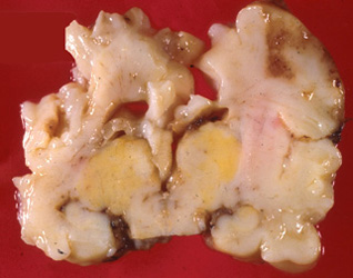
Question 9: This baby was born at 36 weeks of gestation and developed E. Coli sepsis and meningitis.
He died two weeks later. The changes in the brain shown below are due
to:
Correct The lesion represents extreme destruction of brain from HIE,
probably resulting from septic shock. A severe neonatal HSV infection plus HIE may possibly
cause similar lesions.
Incorrect. The lesion represents extreme destruction of brain from HIE,
probably resulting from septic shock. A severe neonatal HSV infection plus HIE
may possibly cause similar lesions.
Incorrect. The lesion represents extreme destruction of brain from
HIE probably resulting from septic shock. A severe neonatal HSV infection plus HIE may possibly cause similar lesions.
Incorrect. The lesion represents extreme destruction of brain from HIE,
probably resulting from septic shock. A severe neonatal HSV infection plus HIE may possibly
cause similar lesions.
Question 10: This child was born at term and had spasticity and psychomotor retardation.
He lived in an institution for retarded children and died at 12 years of age. The brain
lesion shown below is due to:
Incorrect The picture shows a gap in the right hemisphere due to loss of brain tissue.
The left hemisphere had an identical lesion. The pathology is schizencephaly. It is most commonly
a disruption caused by ischemia and other insults occurring in utero. However,
genetic forms of schizencephaly have been described recently.
Correct. The picture shows a gap in the right hemisphere due to loss of brain tissue.
The left hemisphere had an identical lesion. The pathology is schizencephaly.
It is most commonly a disruption caused by ischemia and other insults occurring in utero.
However, genetic forms of schizencephaly have been described recently.
Incorrect. The picture shows a gap in the right hemisphere due to loss of brain tissue.
The left hemisphere had an identical lesion. The pathology is schizencephaly. It is most commonly
a disruption caused by ischemia and other insults occurring in utero.
However, genetic forms of schizencephaly have been described recently.
Question 11
This baby was born at term. He was hypotonic, had a large transilluminating head,
and died at 2 days of age. The brain at autopsy is shown below. Which of the following statements
about the brain pathology is not true:
Correct. The cerebral hemispheres are replaced with a fluid-filled sac.
Small portions of the temporal lobes remain. The cerebellum and brainstem are intact.
The lesion is hydranencephaly, not hydrocephalus. B, C, and D are true of hydranencephaly.
Incorrect.The cerebral hemispheres are replaced with a fluid-filled sac.
Small portions of the temporal lobes remain. The cerebellum and brainstem are intact.
The lesion is hydranencephaly, not hydrocephalus. B, C, and D are true of hydranencephaly.
Incorrect. The cerebral hemispheres are replaced with a fluid-filled sac.
Small portions of the temporal lobes remain. The cerebellum and brainstem are intact.
The lesion is hydranencephaly, not hydrocephalus. B, C, and D are true of hydranencephaly.
Incorrect. The cerebral hemispheres are replaced with a fluid-filled sac.
Small portions of the temporal lobes remain. The cerebellum and brainstem are intact.
The lesion is hydranencephaly, not hydrocephalus. B, C, and D are true of hydranencephaly.
Question 12:
A premature baby developed spastic cerebral palsy. The pathology shown below is due to:
Incorrect. The lesions are cystic PVL. The left image demonstrates classic periventricular
cavitated lesions. The right is a microscopic view of these lesions.
Incorrect. The lesions are cystic PVL. The left image demonstrates classic periventricular cavitated lesions.
The right is a microscopic view of these lesions.
Incorrect. The lesions are cystic PVL. The left image demonstrates classic periventricular cavitated lesions.
The right is a microscopic view of these lesions.
Correct. The lesions are cystic PVL. The left image demonstrates classic periventricular cavitated lesions.
The right is a microscopic view of these lesions
Question 13:
This baby was born at 28 weeks of gestation. At 24 hours, he became hypotonic and his hematocrit dropped to 23.
He died three days later. The pathology illustrated below is due to:
Incorrect. The picture shows blood in the 4th ventricle and around its exit foramina, which is the result of germinal matrix hemorrhage with ventricular rupture (IVH).
Correct. The picture shows blood in the 4th ventricle and around its exit foramina, which is the result of germinal matrix hemorrhage with ventricular rupture (IVH).
image.
Incorrect. The picture shows blood in the 4th ventricle and around its exit foramina, which is the result of germinal matrix hemorrhage with ventricular rupture (IVH).
Incorrect. The picture shows blood in the 4th ventricle and around its exit foramina, which is the result of germinal matrix hemorrhage with ventricular rupture (IVH).
Question 14:
The pathology in the brain of this six day old girl shown below may be caused
by:
Correct. The image shows bilirubin encephalopathy.
This is caused by unconjugated hyperbilirubinemia, which can result from glucuronyl transferase deficiency.
Incorrect. The image shows bilirubin encephalopathy.
This is caused by unconjugated hyperbilirubinemia, which can result from glucuronyl transferase deficiency.
Incorrect. The image shows bilirubin encephalopathy.
This is caused by unconjugated hyperbilirubinemia, which can result from glucuronyl transferase deficiency.
Incorrect. The image shows bilirubin encephalopathy.
This is caused by unconjugated hyperbilirubinemia, which can result from glucuronyl transferase deficiency.