Question 1: A natural epidural space exists around
Incorrect. A natural epidural space occurs only around the spinal cord.
In the cranium, the dura is attached to the inner table of the skull.
Correct. A natural epidural space occurs only around the spinal cord.
In the cranium, the dura is attached to the inner table of the skull.
Incorrect. A natural epidural space occurs only around the spinal cord.
In the cranium, the dura is attached to the inner table of the skull.
Incorrect. A natural epidural space occurs only around the spinal cord.
In the cranium, the dura is attached to the inner table of the skull.
Question 2: The most common cause of subdural empyema is:
Incorrect. Infection spreads to the subdural space from air sinuses or from the middle ear.
Incorrect. Infection spreads to the subdural space from air sinuses or from the middle ear.
Incorrect. Infection spreads to the subdural space from air sinuses or from the middle ear.
Correct. Infection spreads to the subdural space from air sinuses or from the middle ear.
Question 3: A 50 year old patient was admitted to the hospital with a hemorrhagic rash, a temperature of 40C, and shock. He was treated with antibiotics but died in 3 hours.
The best way establish the diagnosis in this case is:
Incorrect. The history is most consistent with Gram negative sepsis. Cultures would be negative because of antibiotics. Latex study of blood or CSF for Neisseria meninginitis is most likely to establish the diagnosis.
Incorrect. The history is most consistent with Gram negative sepsis. Cultures would be negative because of antibiotics. Latex study of blood or CSF for Neisseria meninginitis is most likely to establish the diagnosis.
Correct. The history is most consistent with Gram negative sepsis. Cultures would be negative because of antibiotics. Latex study of blood or CSF for Neisseria meninginitis is most likely to establish the diagnosis.
Incorrect. The history is most consistent with Gram negative sepsis. Cultures would be negative because of antibiotics. Latex study of blood or CSF for Neisseria meninginitis is most likely to establish the diagnosis.
Question 4: Which is more likely to develop one week after the onset of untreated bacterial meningitis?
Correct. Ischemic infarcts may develop in a few days because of vasculitis. Hypoxic-ischemic encephalopathy may also develop, usually earlier. Deafness and hydrocephalus develop much later.
Incorrect. Ischemic infarcts may develop in a few days because of vasculitis. Hypoxic-ischemic encephalopathy may also develop, usually earlier. Deafness and hydrocephalus develop much later.
Incorrect. Ischemic infarcts may develop in a few days because of vasculitis. Hypoxic-ischemic encephalopathy may also develop, usually earlier. Deafness and hydrocephalus develop much later.
Incorrect. Ischemic infarcts may develop in a few days because of vasculitis. Hypoxic-ischemic encephalopathy may also develop, usually earlier. Deafness and hydrocephalus develop much later.
Question 5: The most dangerous feature of an abscess is:
Incorrect. The most life-threatening feature of an abscess is cerebral edema and increased intracranial pressure.
Incorrect. The most life-threatening feature of an abscess is cerebral edema and increased intracranial pressure.
Correct. The most life-threatening feature of an abscess is cerebral edema and increased intracranial pressure.
Incorrect. The most life-threatening feature of an abscess is cerebral edema and increased intracranial pressure.
Question 6: Which of the following is not seen in CNS syphilis ?
Incorrect.
Correct.
Incorrect.
Incorrect.
Question 7: A 3 year old boy presents to the ER in mid September with a history of diarrhea, headache and obtundation. A spinal tap is done. The most appropriate studies are:
Correct. The history is consistent with enterovirus infection. RT PCR will be diagnostic but bacterial cultures and PCR for HSV are usually ordered on the same CSF specimen.
Incorrect. The history is consistent with enterovirus infection. RT PCR will be diagnostic but bacterial cultures and PCR for HSV are usually ordered on the same CSF specimen.
Incorrect. The history is consistent with enterovirus infection. RT PCR will be diagnostic but bacterial cultures and PCR for HSV are usually ordered on the same CSF specimen.
Incorrect. The history is consistent with enterovirus infection. RT PCR will be diagnostic but bacterial cultures and PCR for HSV are usually ordered on the same CSF specimen.
Question 8: Familial CJD cases are:
Correct.
Incorrect.
Incorrect.
Question 9: Most CJD cases are:
Incorrect.
Correct.
Incorrect.
Incorrect.
Question 10: Are there prion protein gene mutations in sporadic CJD?
Incorrect. No prion protein mutations are seen in sporadic CJD. However, the PrP gene shows a polymorphism at codon 129 that can code for either Methionine or Valine. Most patients with sporadic and transmitted CJD are homozygotes for Methionine.
Correct. No prion protein mutations are seen in sporadic CJD. However, the PrP gene shows a polymorphism at codon 129 that can code for either Methionine or Valine. Most patients with sporadic and transmitted CJD are homozygotes for Methionine.
Question 11: Meningitis usually crosses the pial barrier and involves the brain
Incorrect. The pia is a very tough structural barrier. It is not easily trespassed and helps confine meningitis to the subarachnoid space.
Correct. The pia is a very tough structural barrier. It is not easily trespassed and helps confine meningitis to the subarachnoid space.
Question 12: A 15 year old boy was admitted to the hospital with a hemorrhagic rash, temperature of 40C and shock. He was treated with antibiotics but died 5 hours later. The autopsy showed:
Incorrect. The history is consistent with Gram negative shock.
The autopsy usually reveals evidence of DIC and adrenal hemorrhage. Five hours is too soon for meningitis or other inflammatory manifestation to develop.
Incorrect. The history is consistent with Gram negative shock.
The autopsy usually reveals evidence of DIC and adrenal hemorrhage. Five hours is too soon for meningitis or other inflammatory manifestation to develop.
Correct. The history is consistent with Gram negative shock.
The autopsy usually reveals evidence of DIC and adrenal hemorrhage. Five hours is too soon for meningitis or other inflammatory manifestation to develop.
Incorrect. The history is consistent with Gram negative shock.
The autopsy usually reveals evidence of DIC and adrenal hemorrhage. Five hours is too soon for meningitis or other inflammatory manifestation to develop.
Question 13: A 36 year old AIDS patient had fever, headaches, and neurological deficits. MRI showed multiple enhancing brain lesions.
A stereotactic biopsy of one lesion was done. The findings are consistent with:
Incorrect. The picture shows a toxoplasma cyst.
Toxoplasmosis is one of the most common opportunistic infections in AIDS.
Correct. The image shows a toxoplasma cyst.
Toxoplasmosis is one of the most common opportunistic infections in AIDS.
Incorrect. The image shows a toxoplasma cyst.
Toxoplasmosis is one of the most common opportunistic infections in AIDS.
Incorrect. The image shows a toxoplasma cyst.
Toxoplasmosis is one of the most common opportunistic infections in AIDS.
Question 14: *A 6 day old baby developed sepsis, hypotonia, respiratory insufficiency, and died.
The autopsy revealed the brain stem (left) and spinal cord (right) lesions shown below.
The most likely diagnosis is:
Incorrect. Both pictures show microglial nodules among large pyramidal neurons (anterior horns). The location of the lesions is most consistent with enterovirus (Coxsackie) meningoencephalitis. CMV, HSV, and toxoplasma may affect the spinal cord but this is very rare.
Incorrect. Both pictures show microglial nodules among large
pyramidal neurons (anterior horns). The location of the lesions is most
consistent with enterovirus (Coxsackie) meningoencephalitis. CMV, HSV, and toxoplasma may affect the spinal cord but this is very rare.
Correct. Both pictures show microglial nodules among large
pyramidal neurons (anterior horns). The location of the lesions is most
consistent with enterovirus (Coxsackie) meningoencephalitis. CMV, HSV, and toxoplasma may affect the spinal cord but this is very rare.
Incorrect. Both pictures show microglial nodules among large
pyramidal neurons (anterior horns). The location of the lesions is most consistent
with enterovirus (Coxsackie) meningoencephalitis. CMV, HSV, and toxoplasma may affect the spinal cord but this is very rare.
Question 15: The lesion illustrated below may be caused by:
Incorrect. The image shows a multinucleated giant cell without epithelioid cell granulomas.
The lesion is most characteristic of HIV encephalitis.
Correct. The image shows a multinucleated giant cell without epithelioid cell granulomas.
The lesion is most characteristic of HIV encephalitis.
Incorrect. The image shows a multinucleated giant cell without epithelioid cell granulomas.
The lesion is most characteristic of HIV encephalitis.
Incorrect. The image shows a multinucleated giant cell without epithelioid cell granulomas.
The lesion is most characteristic of HIV encephalitis.
Question 16: The pathology shown below most likely represents:
Incorrect. The pathology illustrated is spongiosis of the cortex, characteristic of CJD.
There are also degenerated (pycnotic) neurons. The lesions of Vitamin B12 deficiency involve the spinal cord and those of CO poisoning involve the white matter.
Incorrect. The pathology illustrated is spongiosis of the cortex, characteristic of CJD.
There are also degenerated (pycnotic) neurons. The lesions of Vitamin B12 deficiency involve the spinal cord and those of CO poisoning involve the white matter.
Correct. The pathology illustrated is spongiosis of the cortex, characteristic of CJD.
There are also degenerated (pycnotic) neurons. The lesions of Vitamin B12 deficiency involve the spinal cord and those of CO poisoning involve the white matter.
Incorrect. The pathology illustrated is spongiosis of the cortex, characteristic of CJD.
There are also degenerated (pycnotic) neurons. The lesions of Vitamin B12 deficiency involve the spinal cord and those of CO poisoning involve the white matter.
Question 17: Animal prion diseases occur naturally in the US
Correct. Animal prion diseases occur naturally in elk and mink.
Incorrect. Animal prion diseases occur naturally in elk and mink.
Question 18: The CSF in a patient with the lesion illustrated below shows:
Incorrect. The lesion illustrated is a necrotic mass lesion with numerous giant cells, consistent with a tuberculoma. The CSF shows mononuclear pleocytosis, increased protein and decreased glucose.
Incorrect. The lesion illustrated is a necrotic mass lesion with numerous giant cells, consistent with a tuberculoma. The CSF shows mononuclear pleocytosis, increased protein and decreased glucose.
Correct. The lesion illustrated is a necrotic mass lesion with numerous giant cells, consistent with a tuberculoma. The CSF shows mononuclear pleocytosis, increased protein and decreased glucose.
Incorrect. The lesion illustrated is a necrotic mass lesion with numerous giant cells, consistent with a tuberculoma. The CSF shows mononuclear pleocytosis, increased protein and decreased glucose.
Question 19: The lesions illustrated below are caused by:
Incorrect. The cystic lesions in the cerebellum and cerebrum are consistent with cryptococcal meningitis. The organism is a yeast.
Correct. The cystic lesions in the cerebellum
and cerebrum are consistent with cryptococcal meningitis.
The organism is a yeast.
Incorrect. The cystic lesions in the cerebellum and cerebrum are consistent with cryptococcal meningitis. The organism is a yeast.
Incorrect. The cystic lesions in the cerebellum and cerebrum are consistent with cryptococcal meningitis. The organism is a yeast.
Question 20: The lesion illustrated below is caused by:
Correct. The image shows an intranuclear inclusion which occurs primarily in DNA viruses (such as HSV, CMV, VZV, Papova) and some RNA viruses like measles.
Incorrect. The image shows an intranuclear inclusion which occurs primarily in DNA viruses (such as HSV, CMV, VZV, Papova) and some RNA viruses like measles.
Incorrect. The image shows an intranuclear inclusion which occurs primarily in DNA viruses (such as HSV, CMV, VZV, Papova) and some RNA viruses like measles.
Incorrect. The image shows an intranuclear inclusion which occurs primarily in DNA viruses (such as HSV, CMV, VZV, Papova) and some RNA viruses like measles.
Question 21: The lesion illustrated below is caused by:
Incorrect. The illustrated pathology, necrosis and punctate hemorrhages in the inferior frontal and temporal lobes, is most commonly caused by HSV encephalitis.
Incorrect. The illustrated pathology, necrosis and punctate hemorrhages in the inferior frontal and temporal lobes, is most commonly caused by HSV encephalitis.
Correct. The illustrated pathology, necrosis and punctate hemorrhages in the inferior frontal and temporal lobes, is most commonly caused by HSV encephalitis.
Incorrect. The illustrated pathology, necrosis and punctate hemorrhages in the inferior frontal and temporal lobes, is most commonly caused by HSV encephalitis.
Question 22: A 60 year old patient presented with fever and headaches for 2 days, seizures and hemorrhagic CSF. MRI showed a left frontotemporal lesion with edema. The biopsy of the hemorrhagic lesion is shown below. The diagnosis can be best obtained by:
Incorrect. The clinical and radiologic findings are consistent with HSV encephalitis. The biopsy shows hemorrhage and perivascular inflammation. HSV PCR will provide the fastest diagnosis.
Correct. The clinical and radiologic findings are consistent with HSV encephalitis. The biopsy shows hemorrhage and perivascular inflammation. HSV PCR will provide the fastest diagnosis.
Incorrect. The clinical and radiologic findings are consistent with HSV encephalitis. The biopsy shows hemorrhage and perivascular inflammation. HSV PCR will provide the fastest diagnosis.
Incorrect. The clinical and radiologic findings are consistent with HSV encephalitis. The biopsy shows hemorrhage and perivascular inflammation. HSV PCR will provide the fastest diagnosis.
Question 23: The lesions shown below are caused by:
Incorrect. The picture shows periventricular and cortical calcifications, most consistent with congenital CMV.
Incorrect. The picture shows periventricular and cortical calcifications, most consistent with congenital CMV.
Correct. The picture shows periventricular and cortical calcifications, most consistent with congenital CMV.
Incorrect. The picture shows periventricular and cortical calcifications, most consistent with congenital CMV.
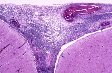
Question 24: The most likely cause of the pathology in the 59 year old patient illustrated below is:
Incorrect. The illustrated lesion is meningitis. The most likely agent in a 59 year old patient is pneumococcus.
Correct. The illustrated lesion is meningitis. The most likely agent in a 59 year old patient is pneumococcus.
Incorrect. The illustrated lesion is meningitis. The most likely agent in a 59 year old patient is pneumococcus.
Incorrect. The illustrated lesion is meningitis. The most likely agent in a 59 year old patient is pneumococcus.