Question 1: What is the most common cause of cerebral ischemia?
Correct Atherosclerosis is indeed the most common cause of cerebral ischemia,
leading to narrowing or occlusion of cerebral arteries.
Incorrect. While hypertension is a major risk factor for stroke,
it is not the most common direct cause of cerebral ischemia.
Incorrect. Cardiac embolism is a significant
cause of stroke
but ranks second to atherosclerosis in frequency.
Incorrect. Vasculitis is a much less common cause of cerebral ischemia compared to
atherosclerosis.
Question 2: If circulation ceases, the energy supplies stored in brain cells are enough to last:
Correct! Energy supplies last for 2 minutes
Incorrect. Energy supplies last for 2 minutes
Incorrect. Energy supplies last for 2 minutes
Incorrect. Energy supplies last for 2 minutes.
Question 3: Neurons damaged by hypoxia or trauma discharge:
Incorrect. Injured neurons discharge glutamate which acts on the same neurons
(autoexcitotoxin) as well as adjacent ones, initiating a destructive cascade.
Correct. Injured neurons discharge glutamate which acts on the same neurons
(autoexcitotoxin) as well as adjacent ones, initiating a destructive cascade.
Incorrect. Injured neurons discharge glutamate which acts on the same neurons
(autoexcitotoxin) as well as adjacent ones, initiating a destructive cascade.
Incorrect. Injured neurons discharge glutamate which acts on the same neurons
(autoexcitotoxin) as well as adjacent ones, initiating a destructive cascade.
Question 4: Free radicals are generated in the:
Incorrect. Free radicals are generated in mitochondria,
the power producing organelles of neurons and all cells.
Incorrect. Free radicals are generated in mitochondria,
the power producing organelles of neurons and all cells.
Correct. Free radicals are generated in mitochondria,
the power producing organelles of neurons and all cells.
Incorrect. Free radicals are generated in mitochondria,
the power producing organelles of neurons and all cells.
Question 5: Which of the following is most vulnerable in HIE?
Incorrect. The caudate nucleus is the most sensitive among the listed structures in
adult HIE.
Correct. The caudate nucleus is the most sensitive among the listed structures in
adult HIE.
Incorrect. The caudate nucleus is the most sensitive among the listed structures in
adult HIE.
Incorrect. The caudate nucleus is the most sensitive among the listed structures in
adult HIE.
Question 6: Cerebral edema in HIE is caused by:
Incorrect. Cerebral edema in HIE is caused by both,
arachidonic acid and lactic acid. Arachidonic acid increases vascular permeability and lactic
acid damages
blood vessels and acts as an osmotic agent.
Incorrect. Cerebral edema in HIE is caused by both, arachidonic acid and lactic
acid.
Arachidonic acid increases vascular permeability
and lactic acid damages blood vessels and acts as an osmotic agent.
Correct! Cerebral edema in HIE is caused by both, arachidonic acid and lactic
acid.
Arachidonic acid increases vascular permeability
and lactic acid damages blood vessels and acts as an osmotic agent.
Incorrect. Cerebral edema in HIE is caused by both, arachidonic acid and lactic
acid.
Arachidonic acid increases vascular permeability
and lactic acid damages blood vessels and acts as an osmotic agent.
Question 7: The respirator brain is caused by:
Incorrect. The respirator
brain is due to autolysis of a non-perfused brain.
Correct. The
respirator brain is due to autolysis of a non-perfused brain.
Incorrect. The respirator
brain is due to autolysis of a non-perfused brain.
Incorrect. The respirator brain
is due to autolysis of a non-perfused brain.
Question 8: Most deaths following MCA occlusion in older patients occur:
Incorrect. Most deaths from large MCA infarcts
occur in the first week but usually not during the first day. At that time, the infarct may
still be evolving.
The cause of death is cerebral edema which is not yet full blown on the first day.
Correct. Most deaths from large MCA infarcts
occur in the first week but usually not during the first day. At that time, the infarct may
still be evolving.
The cause of death is cerebral edema which is not yet full blown on the first day.
Incorrect. Most deaths from large MCA infarcts
occur in the first week but usually not during the first day. At that time, the infarct may
still be evolving.
The cause of death is cerebral edema which is not yet full blown on the first day.
Incorrect. Most deaths from large MCA infarcts
occur in the first week but usually not during the first day. At that time, the infarct may
still be evolving.
The cause of death is cerebral edema which is not yet full blown on the first day.
Question 9: Restoring circulation to the ischemic penumbra can limit brain damage in an ischemic
infarct. The window of opportunity for rescuing the penumbra is:
Incorrect! The window of opportunity
for rescuing the ischemic penumbra is 3-4 hours.
Correct! The window of opportunity for rescuing
the ischemic penumbra is 3-4 hours.
Incorrect! The window of opportunity
for rescuing the ischemic penumbra is 3-4 hours.
Incorrect! The window of opportunity
for rescuing the ischemic penumbra is 3-4 hours.
Question 10: Fusiform aneurysms of the basilar artery usually cause:
Correct. Fusiform aneurysms of the basilar artery
usually undergo thrombosis causing ischemic infarction of the pons.
Rupture and hemorrhage is uncommon.
Incorrect. Fusiform aneurysms of the basilar artery
usually undergo thrombosis causing ischemic infarction of the pons.
Rupture and hemorrhage is uncommon.
Incorrect. Fusiform aneurysms of the basilar artery
usually undergo thrombosis causing ischemic infarction of the pons.
Rupture and hemorrhage is uncommon.
Incorrect. Fusiform aneurysms of the basilar artery
usually undergo thrombosis causing ischemic infarction of the pons.
Rupture and hemorrhage is uncommon.
Question 11: Amnesia involving recent and old memory may result from bilateral lesions of:
Incorrect! Amnesia in the Wernicke-Korsakoff Syndrome may occur as a result
of bilateral damage of hippocampus and amygdala or the thalamus.
Incorrect. Amnesia in the Wernicke-Korsakoff Syndrome may occur as a result
of bilateral damage of hippocampus and amygdala or the thalamus.
Correct. Amnesia in the Wernicke-Korsakoff Syndrome may occur as a result
of bilateral damage of hippocampus and amygdala or the thalamus.
Incorrect. Amnesia in the Wernicke-Korsakoff Syndrome may occur as a result
of bilateral damage of hippocampus and amygdala or the thalamus.
Question 12: The intracellular process that triggers cell injury in HIE is:
Correct. Increased intracellular calcium activates proteases, phospholipases,
and endonucleases that cause cellular destruction.
Incorrect. Increased intracellular calcium activates proteases, phospholipases,
and endonucleases that cause cellular destruction.
Incorrect. Increased intracellular calcium activates proteases, phospholipases,
and endonucleases that cause cellular destruction.
Incorrect. Increased intracellular calcium activates proteases, phospholipases,
and endonucleases that cause cellular destruction.
Question 13: The persistent vegetative state may result from extensive damage of:
Incorrect! The persistent vegetative state results
from extensive damage of either the cerebral cortex or the thalamus, or both.
Correct. The persistent vegetative state results from
extensive damage of either the cerebral cortex or the thalamus, or both.
Incorrect. The persistent vegetative state results from
extensive damage of either the cerebral cortex or the thalamus, or both.
Incorrect. The persistent vegetative state results from
extensive damage of either the cerebral cortex or the thalamus, or both.
Question 14: Intracranial arterial aneurysms can cause all of the following except:
Correct. Pontine hemorrhage is a parenchymal hemorrhage
usually caused by hypertension. Aneurysmal bleeds can result in parenchymal hemorhage in some
cases, such hemorrhage usually involves the cerebrum. Large aneurysms can compress cranial
nerves. Hydrocephalus may develop after subarachnoid hemorrhage, due to meningeal fibrosis.
Incorrect. Pontine hemorrhage is a parenchymal hemorrhage usually caused by
hypertension. Aneurysmal bleeds can result in parenchymal hemorhage in some cases, such
hemorrhage usually involves the cerebrum. Large aneurysms can compress cranial nerves.
Hydrocephalus may develop after subarachnoid hemorrhage, due to meningeal fibrosis.
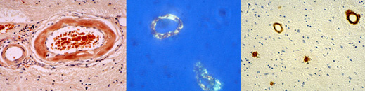
Question 15:The images of cerebral vessels below (Congo Red stain,
left, Congo Red fluorescence, middle and unknown immunostain, right)
illustrate a pathological process associated with:
Incorrect! The images demonstrate vascular amyloid and the immunostain
is beta amyloid. Beta amyloid deposits in the brain occur in Alzheimer’s disease
and can cause also amyloid angiopathy which is associated with lobar intracerebral hemorrhages.
Amyloid deposits are also seen in familial amyloid neuropathy but these are not vacular
and they are not beta amyloid
Incorrect. The images demonstrate vascular amyloid and the immunostain
is beta amyloid. Beta amyloid deposits in the brain occur in Alzheimer’s disease and
can cause also amyloid angiopathy which is associated with lobar intracerebral hemorrhages.
Amyloid deposits are also seen in familial amyloid neuropathy but these are not vascular
and they are not beta amyloid.
Correct. The images demonstrate vascular amyloid and the immunostain
is beta amyloid. Beta amyloid deposits in the brain occur in Alzheimer’s disease and can
cause also amyloid angiopathy which is associated with lobar intracerebral hemorrhages.
Amyloid deposits are also seen in familial amyloid neuropathy but these are not vacular
and they are not beta amyloid.
Incorrect. The images demonstrate vascular amyloid and the immunostain
is beta amyloid. Beta amyloid deposits in the brain occur in Alzheimer’s disease and
can cause also amyloid angiopathy which is associated with lobar intracerebral
hemorrhages.
Amyloid deposits are also seen in familial amyloid neuropathy but these are not vacular
and
they are not beta amyloid.
Question 16: *Risk factors for cerebral arterial occlusion
and ischemic infarction include:
Incorrect. Although factor V Leiden more frequently causes venous
thrombosis,
it can also be associated with arterial thrombosis and ischemic infarction.
Incorrect. Although factor V Leiden more frequently causes venous
thrombosis,
it can also be associated with arterial thrombosis and ischemic infarction.
Correct. Although factor V Leiden more frequently causes venous thrombosis,
it can also be associated with arterial thrombosis and ischemic infarction.
Incorrect. Although factor V Leiden more frequently causes venous
thrombosis,
it can also be associated with arterial thrombosis and ischemic infarction.
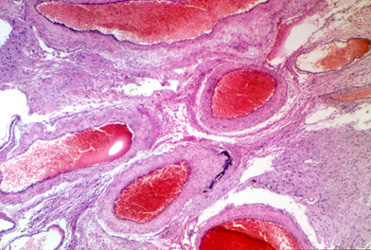
Question 17. The lesion illustrated below can cause:
Incorrect! The illustrated lesion is an AVM. AVMs can cause headaches
due to recurrent bleeds, focal neurologic deficits, and seizures.
Incorrect. The illustrated lesion is an AVM. AVMs can cause headaches
due to recurrent bleeds, focal neurologic deficits, and seizures.
Incorrect. The illustrated lesion is an AVM. AVMs can cause headaches due
to recurrent bleeds, focal neurologic deficits, and seizures.
Correct. The illustrated lesion is an AVM. AVMs can cause
headaches due to recurrent bleeds, focal neurologic deficits, and seizures.
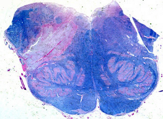
Question 18: The lesion illustrated below may be caused by occlusion of:
Incorrect. The picture shows a section of medulla stained with a myelin stain.
The unstained wedge on the left represents a lateral medullary infarct. It is caused by occlusion
of the posterior inferior cerebellar artery (PICA), which may be a branch of either the vertebral or the basilar artery.
Incorrect. The picture shows a section of medulla stained with a myelin stain.
The unstained wedge on the left represents a lateral medullary infarct. It is caused by occlu
sion of the posterior inferior cerebellar artery (PICA), which may be a branch of either the
vertebral or the basilar artery.
Correct. The picture shows
a section of medulla stained with a myelin stain.The unstained wedge on the left represents
a lateral medullary infarct. It is caused by occlusion of the posterior inferior cerebellar artery (PICA),
which may be a branch of either the vertebral or the basilar artery.
Incorrect. The picture shows a section of medulla stained with a myelin stain.
The unstained wedge on the left represents a lateral medullary infarct. It is caused by occlusion
of the posterior inferior cerebellar artery (PICA),
which may be a branch of either the vertebral or the basilar artery.
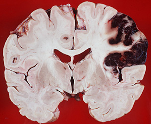
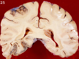
Question 19: The lesion illustrated below is due primarily to vascular occlusion
True. The lesion is a hemorrhagic infarct in the distribution of the RMCA.
The basic mechanism is arterial occlusion, usually by an embolus, with reperfusion and leakage
through a damaged capillary bed following lysis of the embolus.
False. The lesion is a hemorrhagic infarct in the distribution of the RMCA.
The basic mechanism is arterial occlusion, usually by an embolus, with reperfusion and leakage
through a damaged capillary bed following lysis of the embolus.
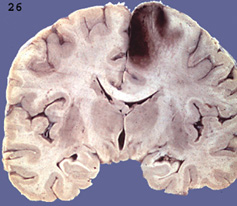
Question 20: The lesion illustrated below can cause severe neurologic deficits
but is no threat to life
Correct. The lesion is a lacunar infarct. Because of its small volume,
it does not cause life threatening cerebral edema but because it is
frequently located in deep structures (internal capsule basal ganglisa brainstem)
it can cause severe neurologic deficits.
Incorrect. The lesion is a lacunar infarct. Because of its small
volume, it does not cause life threatening cerebral edema but because it is frequently
located in deep structures (internal capsule basal ganglisa brainstem)
it can cause severe neurologic deficits.
Question 21: The illustrated lesion is most likely caused by:
Correct. The lesion is a hemorrhagic infarct in the ACA territory,
most likely caused by embolism.
Incorrect. The lesion is a hemorrhagic infarct in the ACA territory, most likely caused by embolism.
Incorrect. The lesion is a hemorrhagic infarct in the ACA territory, most likely caused by embolism.
Incorrect. The lesion is a hemorrhagic infarct in the ACA territory,
most likely caused by embolism.
Question : The lesion shown below is in the territory of the:
Incorrect! The lesion is in the PCA territory.
Incorrect. The lesion is in the PCA territory
Correct. The lesion is in the Posterior Cerebral Artery (PCA) territory.
Incorrect. The lesion is in the PCA territory.
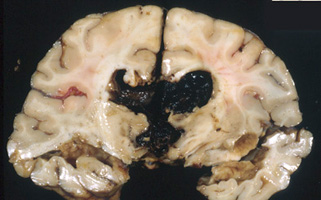
Question 23: The lesion illustrated below is caused by:
Incorrect! Hemorrhage in the third and lateral ventricles resulting
from rupture of an ACA aneurysm.
Incorrect. Hemorrhage in the third and lateral ventricles
resulting from rupture of an ACA aneurysm.
Incorrect. Hemorrhage in the third
and lateral ventricles resulting from rupture of an ACA aneurysm.
Correct. Hemorrhage in the third and lateral ventricles
resulting from rupture of an ACA aneurysm.
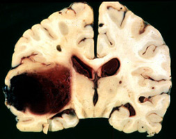
Question 24: A 72 year old woman with a one year history of declining memory developed sudden headache
and decreased consciousness and collapsed while washing dishes. Neuropathological examination revealed the lesion below.
What is the most likely cause of the lesion?
Incorrect! The lesion is a lobar hemorrhage. The cause, in this case, was cerebral amyloid angiopathy.
Patients with CAA may have Alzheimer disease.
Hypertension can cause similar lesions. Anticoagulation can cause a variety of hemorrhages,
usually associated with head trauma.rebral arteries.
Correct. The lesion is a lobar hemorrhage. The cause, in this case, was cerebral amyloid angiopathy.
Patients with CAA may have Alzheimer disease.
Hypertension can cause similar lesions. Anticoagulation can cause a variety of hemorrhages,
usually associated with head trauma.
Incorrect. The lesion is a lobar hemorrhage. The cause, in this case,
was cerebral amyloid angiopathy. Patients with CAA may have Alzheimer disease.
Hypertension can cause similar lesions. Anticoagulation can cause a variety of hemorrhages, usually associated with head trauma.
Incorrect. The lesion is a lobar hemorrhage. The cause, in this case, was cerebral amyloid angiopathy.
Patients with CAA may have Alzheimer disease. Hypertension can cause similar lesions. Anticoagulation can cause a variety of hemorrhages, usually associated with head trauma.
ypertension can cause similar lesions. Anticoagulation can cause a variety of hemorrhages, usually associated with head trauma.
Question 25: A 15 year old male with SLE had decreased consciousness, bilateral lower extremity weakness,
and the MRI changes illustrated below.
Symptoms improved somewhat but a follow-up MRI showed residual encephalomalacia. The lesions are most likely caused by:
Incorrect! The T2 MRI shows bilateral parasagittal lesions, consistent with venous infarcts.
The most likely etiology is superior sagittal sinus thrombosis associated with the anticardiolipin syndrome.
Incorrect. The T2 MRI shows bilateral parasagittal lesions, consistent with venous infarcts.
The most likely etiology is superior sagittal sinus thrombosis associated with the anticardiolipin syndrome.
Incorrect. The T2 MRI shows bilateral parasagittal lesions,
consistent with venous infarcts. The most likely etiology is superior sagittal sinus thrombosis
associated with the anticardiolipin syndrome.
Correct. The T2 MRI shows bilateral parasagittal lesions, consistent with venous infarcts.
The most likely etiology is superior sagittal sinus thrombosis associated with the anticardiolipin syndrome.
Question 26: The MRI images shown below were obtained one month apart (the left first).
The illustrated pathology can cause:
Incorrect! The picture on the right shows bilateralhippocampal atrophy,
indicated by smaller hippocampi and more ample temporal horns of of the lateral ventricles. of the lateral ventricles.
Incorrect. The lesion is hippocampal sclerosis and can cause seizures and Korsakoff amnesia.
The image on the right shows smaller hippocampi and more empty space in the temporal horns of the lateral ventricles.
Correct. The lesion is hippocampal sclerosis and can cause
seizures and Korsakoff amnesia. The image on the right shows smaller hippocampi and more empty space in the
temporal horns of the lateral ventricles.
Incorrect. The lesion is hippocampal sclerosis and can cause seizures and Korsakoff amnesia.
The image on the right shows smaller hippocampi and more empty space in the temporal horns of the lateral ventricles.