Question 1: Ischemic lesions or HIE are a major component of the shaken baby syndrome
Correct
Incorrect.
Question 2: Which of the following identifies damaged axons in diffuse axonal injury?
Incorrect. The axonal swellings can be identified with immunostains for beta amyloid precursor protein.
Correct The axonal swellings can be identified with immunostains for beta amyloid precursor protein.
Question 3: A fibrous inner membrane encapsulating the subdural hematoma develops in:
Incorrect. A fibrous inner membrane encapsulating the subdural develops in 4-6 weeks.
Incorrect. A fibrous inner membrane encapsulating the subdural develops in 4-6 weeks.
Incorrect. A fibrous inner membrane encapsulating the subdural develops in 4-6 weeks.
Correct. A fibrous inner membrane encapsulating the subdural develops in 4-6 weeks.
Question 4: Pure diffuse axonal injury is characterized by severe brain swelling
Incorrect. No brain swelling is seen in pure diffuse axonal injury.
Correct. No brain swelling is seen in pure diffuse axonal injury.
Question 5: All subdural hematomas have a free interval between the trauma and onset of symptoms
Incorrect. Some subdural hematomas do not
have a free interval between the trauma and onset of symptoms.
Correct. Some subdural hematomas not have a free interval between the trauma and onset of symptoms.
Question 6: Epidural hematomas may occur without a skull fracture
Correct. Epidural hematomas may occur without skull fractures.
The inner plate may deform enough to tear a blood vessel without necessarily breaking.
Most commonly, however, epidural hematomas are seen with skull fractures.
Incorrect. Epidural hematomas may occur without skull fractures.
The inner plate may deform enough to tear a blood vessel without necessarily breaking.
Most commonly, however, epidural hematomas are seen with skull fractures.
Question 7: Transsection of the cervical spinal cord may occur in the shaken baby syndrome
Correct.
Incorrect.
Question 8: Who is more susceptible to developing a subdural hematoma?
Incorrect. Cerebral atrophy stretches bridging veins making an individual more prone to developing subdural hematomas. Thus, a 72 year-old patient with Alzheimer's disease would be more susceptible.
Incorrect. Cerebral atrophy stretches bridging veins making an individual more prone to developing subdural hematomas. Thus, a 72 year-old patient with Alzheimer's disease would be more susceptible.
Incorrect. Cerebral atrophy stretches bridging veins making an individual more prone to developing subdural hematomas. Thus, a 72 year-old patient with Alzheimer's disease would be more susceptible.
Correct Cerebral atrophy stretches bridging veins making an individual more prone to developing subdural hematomas. Thus, a 72 year-old patient with Alzheimer's disease would be more susceptible.
Question 9: Beta amyloid precursor protein is produced by neurons at the time of traumatic brain injury
Incorrect. Beta amyloid precursor protein is normally produced by neurons. It accumulates in damaged axons due to disrupted axonal transport.
Correct. Beta amyloid precursor protein is normally produced by neurons. It accumulates in damaged axons due to disrupted axonal transport.
Question 10: The most common traumatic brain injury is:
Incorrect. Subarachnoid hemorrhage is the most frequent traumatic brain lesion.
Incorrect. Subarachnoid hemorrhage is the most frequent traumatic brain lesion.
Correct. Subarachnoid hemorrhage is the most frequent traumatic brain lesion.
Incorrect. Subarachnoid hemorrhage is the most frequent traumatic brain lesion.
Incorrect. Eventually, the contusion evolves into a yellowish plaque characterized by loss and atrophy of brain tissue, glial scarring, hemosiderin deposition, and loss of axons in the underlying white matter.
Incorrect. Eventually, the contusion evolves into a yellowish plaque characterized by loss and atrophy of brain tissue, glial scarring, hemosiderin deposition, and loss of axons in the underlying white matter.
Correct. Eventually, the contusion evolves into a yellowish plaque characterized by loss and atrophy of brain tissue, glial scarring, hemosiderin deposition, and loss of axons in the underlying white matter.
Incorrect. Eventually, the contusion evolves into a yellowish plaque characterized by loss and atrophy of brain tissue, glial scarring, hemosiderin deposition, and loss of axons in the underlying white matter.
Question 12: Axonal swellings occur mainly in:
Incorrect. Axonal swellings occur in all of the above conditions.
Incorrect. Axonal swellings occur in all of the above conditions.
Incorrect. Axonal swellings occur in all of the above conditions.
Correct. Axonal swellings occur in all of the above conditions.
Question 13: The mechanism by which axonal swellings occur in diffuse axonal injury involves all of the following processes except:
Correct. Axonal swellings do not result from rupture of axons at the time of injury. Deformation of axon at internodes, calcium influx in the axoplasm, and compaction of neurofilaments occur before axons actually break.
Incorrect. Axonal swellings do not result from rupture of axons at the time of injury. Deformation of axon at internodes, calcium influx in the axoplasm, and compaction of neurofilaments occur before axons actually break.
Incorrect. Axonal swellings do not result from rupture of axons at the time of injury. Deformation of axon at internodes, calcium influx in the axoplasm, and compaction of neurofilaments occur before axons actually break.
Incorrect. Axonal swellings do not result from rupture of axons at the time of injury. Deformation of axon at internodes, calcium influx in the axoplasm, and compaction of neurofilaments occur before axons actually break.
Question 14: Among the following structures, petechiae, in diffuse axonal injury, are found most commonly in:
Incorrect. Petechiae, in DAI, occur most commonly in the corpus callosum and dorsolateral brainstem.
Correct. Petechiae, in DAI, occur most commonly in the corpus callosum and dorsolateral brainstem.
Incorrect. Petechiae, in DAI, occur most commonly in the corpus callosum and dorsolateral brainstem.
Incorrect. Petechiae, in DAI, occur most commonly in the corpus callosum and dorsolateral brainstem.
Question 15: Petechiae, in diffuse axonal injury, besides the corpus callosum, are commonly found in which of the following?
Incorrect. Petechiae, in DAI, occur most commonly in the corpus callosum and dorsolateral brainstem.
Incorrect. Petechiae, in DAI, occur most commonly in the corpus callosum and dorsolateral brainstem.
Incorrect. Petechiae, in DAI, occur most commonly in the corpus callosum and dorsolateral brainstem.
Correct. Petechiae, in DAI, occur most commonly in the corpus callosum and dorsolateral brainstem.
Question 16: The Bielschowsky stain shows axonal swellings in:
Incorrect. H&E and silver stains detect axonal swellings in about 15 hours. Beta amyloid precursor immunostains detect them in 2-3 hours.
Incorrect. H&E and silver stains detect axonal swellings in about 15 hours. Beta amyloid precursor immunostains detect them in 2-3 hours.
Correct. H&E and silver stains detect axonal swellings in about 15 hours. Beta amyloid precursor immunostains detect them in 2-3 hours.
Incorrect. H&E and silver stains detect axonal swellings in about 15 hours. Beta amyloid precursor immunostains detect them in 2-3 hours.
Question 17: The brain of a 62 year old former professional boxer who has dementia and Parkinsonian manifestations shows:
Correct. The symptoms, in this setting are consistent with chronic traumatic encephalopathy. Microscopic examination in CTE reveals hyperphosphorylated tau (p-tau) deposition primarily in neurons and around small vessels.
Incorrect. The symptoms, in this setting are consistent with chronic traumatic encephalopathy. Microscopic examination in CTE reveals hyperphosphorylated tau (p-tau) deposition primarily in neurons and around small vessels.
Incorrect. The symptoms, in this setting are consistent with chronic traumatic encephalopathy. Microscopic examination in CTE reveals hyperphosphorylated tau (p-tau) deposition primarily in neurons and around small vessels.
Incorrect. The symptoms, in this setting are consistent with chronic traumatic encephalopathy. Microscopic examination in CTE reveals hyperphosphorylated tau (p-tau) deposition primarily in neurons and around small vessels.
Question 18: A patient with a glioblastoma multiforme in the right frontoparietal area develops right hemiparesis and a fixed dilated right pupil. The cause of these neurological findings is:
Incorrect. The most likely cause is herniation of the right temporal lobe. This damages the ipsilateral third nerve, causing the right fixed dilated pupil, and compresses the left cerebral peduncle, causing the right hemiparesis.
Incorrect. The most likely cause is herniation of the right temporal lobe. This damages the ipsilateral third nerve, causing the right fixed dilated pupil, and compresses the left cerebral peduncle, causing the right hemiparesis.
Correct. The most likely cause is herniation of the right temporal lobe. This damages the ipsilateral third nerve, causing the right fixed dilated pupil, and compresses the left cerebral peduncle, causing the right hemiparesis.
Incorrect. The most likely cause is herniation of the right temporal lobe. This damages the ipsilateral third nerve, causing the right fixed dilated pupil, and compresses the left cerebral peduncle, causing the right hemiparesis.
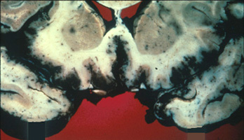
Question 19: The pathology in the section across the midbrain shown below is the result of:
Incorrect. The picture shows herniation of the left temporal lobe (right on the picture). There is displacement of the midbrain, compression of the contralateral cerebral peduncle, and secondary brainstem hemorrhages. The most likely diagnosis is a left subdural hematoma.
Correct. The picture shows herniation of the left temporal lobe (right on the picture). There is displacement of the midbrain, compression of the contralateral cerebral peduncle, and secondary brainstem hemorrhages. The most likely diagnosis is a left subdural hematoma.
Incorrect. The picture shows herniation of the left temporal lobe (right on the picture). There is displacement of the midbrain, compression of the contralateral cerebral peduncle, and secondary brainstem hemorrhages. The most likely diagnosis is a left subdural hematoma.
Incorrect. The picture shows herniation of the left temporal lobe (right on the picture). There is displacement of the midbrain, compression of the contralateral cerebral peduncle, and secondary brainstem hemorrhages. The most likely diagnosis is a left subdural hematoma.
Question 20: The pathological lesions shown below are the result of:
Incorrect. Although theoretically anticoagulant treatment, DAI, and hypertension may cause brainstem hemorrhages, the most common cause of the hemorrhages shown is herniation from increased intracranial pressure (secondary brainstem hemorrhages).
Incorrect. Although theoretically anticoagulant treatment, DAI, and hypertension may cause brainstem hemorrhages, the most common cause of the hemorrhages shown is herniation from increased intracranial pressure (secondary brainstem hemorrhages).
Incorrect. Although theoretically anticoagulant treatment, DAI, and hypertension may cause brainstem hemorrhages, the most common cause of the hemorrhages shown is herniation from increased intracranial pressure (secondary brainstem hemorrhages).
Correct. Although theoretically anticoagulant treatment, DAI, and hypertension may cause brainstem hemorrhages, the most common cause of the hemorrhages shown is herniation from increased intracranial pressure (secondary brainstem hemorrhages).
Question 21: The lesions in this 7 month old baby girl shown below are most likely caused by:
Incorrect. The picture shows hemorrhage around the optic nerve. The most common cause of optic nerve and retinal hemorrhage in a 7 month-old infant is traumatic brain injury, specifically the shaken baby syndrome. However, coagulopathy, resuscitation, and chest trauma have been implicated in rare cases.
Incorrect. The picture shows hemorrhage around the optic nerve. The most common cause of optic nerve and retinal hemorrhage in a 7 month-old infant is traumatic brain injury, specifically the shaken baby syndrome. However, coagulopathy, resuscitation, and chest trauma have been implicated in rare cases.
Incorrect. The picture shows hemorrhage around the optic nerve. The most common cause of optic nerve and retinal hemorrhage in a 7 month-old infant is traumatic brain injury, specifically the shaken baby syndrome. However, coagulopathy, resuscitation, and chest trauma have been implicated in rare cases.
Correct. The picture shows hemorrhage around the optic nerve. The most common cause of optic nerve and retinal hemorrhage in a 7 month-old infant is traumatic brain injury, specifically the shaken baby syndrome. However, coagulopathy, resuscitation, and chest trauma have been implicated in rare cases.
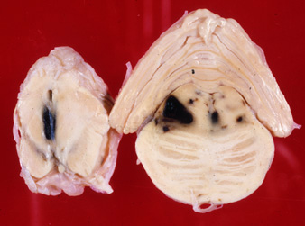
Question 22: The most likely mechanism for the lesions shown below is:
Incorrect. The hemorrhagic white matter lesions are characteristic of gliding contusions, a form of traumatic brain injury caused by excessive movement of the brain inside the skull resulting from blows to the head and other mechanisms.
Incorrect. The hemorrhagic white matter lesions are characteristic of gliding contusions, a form of traumatic brain injury caused by excessive movement of the brain inside the skull resulting from blows to the head and other mechanisms.
Correct. The hemorrhagic white matter lesions are characteristic of gliding contusions, a form of traumatic brain injury caused by excessive movement of the brain inside the skull resulting from blows to the head and other mechanisms.
Incorrect. The hemorrhagic white matter lesions are characteristic of gliding contusions, a form of traumatic brain injury caused by excessive movement of the brain inside the skull resulting from blows to the head and other mechanisms.
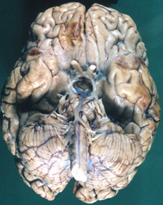
Question 23: The brain lesions in the 57 year old man shown below are most likely due to:
Incorrect. The most likely cause of bilateral hemorrhagic necrosis of the inferior frontal and temporal lobes is HSV encephalitis. However, contre-coup contusions may have similar distribution. PCA occlusion does not involve the frontal lobes.
Correct. The most likely cause of bilateral hemorrhagic necrosis of the inferior frontal and temporal lobes is HSV encephalitis. However, contre-coup contusions may have similar distribution. PCA occlusion does not involve the frontal lobes.
Incorrect. The most likely cause of bilateral hemorrhagic necrosis of the inferior frontal and temporal lobes is HSV encephalitis. However, contre-coup contusions may have similar distribution. PCA occlusion does not involve the frontal lobes.
Incorrect. The most likely cause of bilateral hemorrhagic necrosis of the inferior frontal and temporal lobes is HSV encephalitis. However, contre-coup contusions may have similar distribution. PCA occlusion does not involve the frontal lobes.
Question 24: The lesions illustrated below are most likely to occur as a result of:
Incorrect. The lesions are old contre-coup contusions. While they can occur with a variety of traumatic injuries, they are most common with falls to the back.
Incorrect. The lesions are old contre-coup contusions. While they can occur with a variety of traumatic injuries, they are most common with falls to the back.
Correct. The lesions are old contre-coup contusions. While they can occur with a variety of traumatic injuries, they are most common with falls to the back.
Incorrect. The lesions are old contre-coup contusions. While they can occur with a variety of traumatic injuries, they are most common with falls to the back.
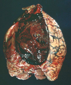
Question 25: The image below shows:
Incorrect. The image shows a subdural hematoma with the dura flipped over.
Correct. The image shows a subdural hematoma with the dura flipped over.
Incorrect. The image shows a subdural hematoma with the dura flipped over.
Incorrect. The image shows a subdural hematoma with the dura flipped over.