M2 NEUROPATHOLOGY LAB
QUIZ 9
Case A
Case B
Case C
Case D
Case E
Match the case histories with images 41-45.
41 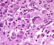 42
42 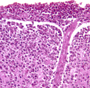 43
43 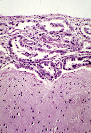 44
44 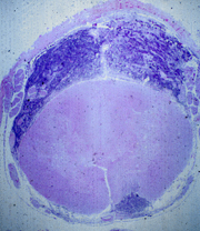 45
45 
Case A
A 70-year-old man had a sudden onset of slurring of
speech and right arm weakness which resolved in 30
minutes. However, right hemiparesis, facial paralysis,
and slurred speech reappeared insidiously within
a few days and progressed. MRI revealed an enhancing
mass involving the left frontal lobe, basal ganglia,
and internal capsule. The patient had AIDS which
he had presumably acquired from a blood transfusion
several years earlier, in the course of partial gastrectomy
for peptic ulcer.
Case B
A 50-year-old man had seizures in the left hand for
several years. Then weakness of the hand and arm
developed, followed by a drop of the left side of
the mouth. Seizures became more frequent and headaches
appeared. Brain MRI revealed a right parasagittal
extra-axial mass which was totally resected, leading
to complete recovery.
Case C
A 74-year-old woman who had a right parietal headache
for one month developed weakness and numbness of
the left hand and began to drop things. The whole
left arm then became weak, and drooping of the left
side of the mouth developed, along with a sensation
of thick tongue. Weakness progressed to involve her
left leg, and a defect in the left visual field appeared.
MRI revealed an enhancing right parietal mass. A
craniotomy was done, and a necrotic tumor was partially
resected.
Case D
A 3 year-old girl had a cerebellar tumor which was
partially resected. She received intrathecal chemotherapy
and craniospinal irradiation and was stable for ten
months. Then, she developed tingling of the toes,
back pain, and weakness in the legs. A lumbar puncture
revealed tumor cells in the CSF. Symptoms progressed,
and within three months she lost sensation below
the chest, and became paraplegic and incontinent.
Additional chemotherapy and irradiation was given,
but she died four months later.
Case E
A 62-year-old woman who had had mastectomy for breast
cancer two years earlier gradually became confused,
ataxic, weak, and dysarthric. Head CT was normal.
CSF was clear with protein 136 mg/dl, glucose 14
mg/dl, and 30 lymphocytes. Her condition worsened,
and a succession of cranial nerve deficits appeared.
She deteriorated further, became comatose, and died
2 ½
months after the onset of the neurological symptoms.
ALL LECTURE PODCASTS ARE EMBEDDED IN THEIR RESPECTIVE
NEUROPATHOLOGY PAGES AND CAN ALSO BE FOUND ON THE
VIMEO CHANNEL "DIMITRI
AGAMANOLIS NEUROPATHOLOGY"