METABOLIC MYOPATHIES
Glycogenosis type V (phosphorylase deficiency, McArdle disease), and type VII (phosphofructokinase deficiency, Tarui disease) are glycolytic enzyme defects that impair the use of glycogen for energy. They cause weakness, cramps, and myonecrosis on exertion. Between episodes, the muscle may look normal. In phosphofructokinase deficiency, myofibers contain insoluble abnormal glycogen with a filamentous fine structure, similar to the long chain unbranched glycogen that is seen in Glycogenosis type IV-Andersen disease. There are specific histochemical and biochemical tests for diagnosis of these glycogenoses.
 Pompe disease |
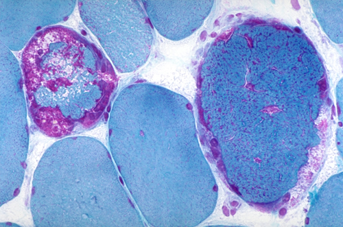
 Pompe disease, skin biopsy |
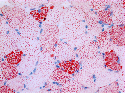
 Adult acid maltase deficiency |
 Lipid storage myopathy |
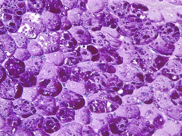
In glycogenosis type II (Pompe disease-acid maltase deficiency), lysosomal storage of glycogen in skeletal and cardiac muscle causes hypotonia, cardiomyopathy, and death in infancy. There is also glycogen storage in the liver, kidneys, brain, and other organs. Milder mutations cause an adult onset vacuolar myopathy.
Disorders of lipid metabolism impair the use of lipid for energy. Carnitine carries fatty acids into the mitochondria. Carnitine deficiency causes weakness and a lipid storage myopathy. Carnitine palmitoyl transferase (CPT) transfers fatty acids from carnitine to acetyl-CoA. CPT deficiency causes myonecrosis, often precipitated by exercise.
 Ragged red fibers |
 Ragged blue fiber |
 COX-negative fibers |
 Mitochondria in RRF |
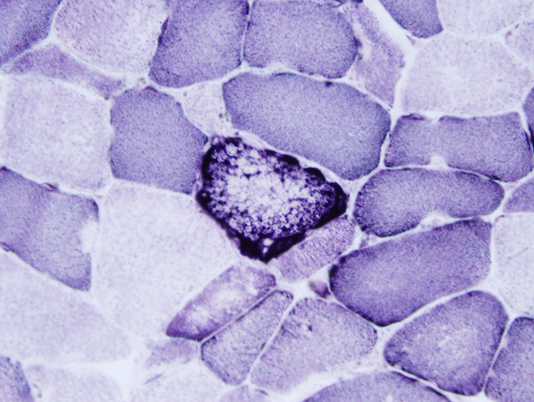
Deficiencies of enzymes of the respiratory chain affect more severely muscle and brain which are heavily dependent on oxidative phosphorylation. Several mitochondrial encephalopathy and encephalomyopathy syndromes have been described. Muscle involvement is characterized by weakness and ophthalmoplegia. Mitochondrial myopathies that impair intramitochondrial protein synthesis are associated with a characteristic histological abormality, the "ragged red fiber" (RRF). This term refers to an abnormality seen in frozen sections of muscle stained with the Gomori trichrome stain. The red color of these fibers is due to large numbers of abnormal mitochondria that represent a compensatory proliferation. In addition to their red color, the abnormal fibers are coarse and disorganized. The same fibers are also called ragged blue fibers because they show an intense, irregular staining with Succinic Dehydrogenase and NADH histochemical stains. Electron microscopy shows clusters of large, irregular mitochondria with crystal-like inclusions. In addition,in some mitochondrial myopathies, especially CPEO, KSS, and MERRF, the muscle contains Cytochrome C Oxidase-negative fibers. Small numbers of COX-negative fibers are also seen in inflammatory myopathies, especially inclusion body myositis, and in old people without muscle disease.
Updated: June, 2014