M2 NEUROPATHOLOGY LAB
QUIZ 10 (microscopic pathology)
Case A
Case B
Case C
Case D
Case E
Match your diagnoses with images 46-50.
46 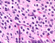 47
47 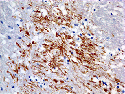 48
48 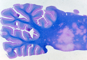 49
49 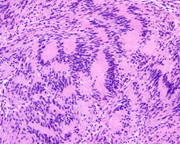 50
50 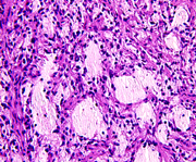
Case A
A 7 year old boy was an unrestrained passenger in
the front seat when the car in which he was riding
was hit by another car on the left side. The air
bag deployed, hitting him in the left temple. In
the ER, he was unconscious, responding only to painful
stimuli by extensor spasms. He had a scalp laceration
and fractures of the skull and left jaw. One week
after the accident, he was still unconscious. His
pupils and eye movements were normal, corneal and
gag reflexes were present, and he had no facial weakness.
He had weakness, spasticity and brisk tendon reflexes
on the right. Both plantar responses were extensor.
He lay most of the time with eyes open and had coordinated
eye movements following sounds and moving objects.
He appeared to sleep at times but could be roused.
He died of pneumonia and sepsis 10 days after the
accident.
Case B
A 4 year old boy had headaches and frequent vomiting
in the morning for 2 weeks. Examination by a pediatrician
and gastroenterology referral revealed no abnormalities.
The symptoms worsened. He became incoordinated and
his vision became blurred. Fundoscopic exam revealed
papilledema. MRI showed a cystic cerebellar mass.
A posterior fossa craniotomy was done and a partly
cystic lesion was removed.
Case C
A 39-year-old woman with Hodgkin’s disease developed
a rapid succession of neurologic deficits. First, there
was left facial weakness, followed by ataxia and weakness
of the left hand. One month later, a left visual field
cut appeared, then left hemiparesis. CSF was normal.
MRI showed areas of demyelination without mass effect.
Two months later, right hemiparesis and upward gaze
paralysis appeared. Gradually she became comatose and
died four months after the onset of her illness.
Case D
A 14 year old boy had buzzing in the left ear
and tingling and pain in the left face for one
year. Loss of hearing and vetigo gradually developed.
Neurological examination revealed weakness and
sensory loss on the left face. MRI revealed a
2.5 cm extra-axial mass in the left cerebellopontine
angle, extending into the left auditory meatus.
His mother had a spinal cord tumor removed when
she was 34 years old. The tumor was removed entirely
through a posterior fossa craniotomy.
Case E
A 61 year old man had a history of focal seizures
involving the left arm for 4 years. Gradually,
weakness and spasticity of the left arm and face
developed. Imaging studies reveiled an isodense
poorly defined, nonenhancing mass in the right
posterior frontal area, above the Sylvian fissure.
The lesion involved cortex and white matter.
Fine calcification was noted on the CT. A biopsy
was done.
ALL LECTURE PODCASTS ARE EMBEDDED IN THEIR RESPECTIVE
NEUROPATHOLOGY PAGES AND CAN ALSO BE FOUND ON THE
VIMEO CHANNEL "DIMITRI
AGAMANOLIS NEUROPATHOLOGY"